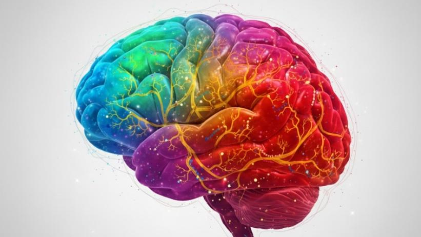
The incredible first map of brain development showing how stem cells transform into neurons.

A new generation of scientific studies is revealing, with greater precision than ever before, how the human brain structures itself as it grows. The efforts of the BRAIN Initiative Cell Atlas Network (BICAN) are shedding light on the enormous diversity of cell types that emerge during brain development in humans, rodents, and non-human primates. These efforts go beyond simply mapping which cells exist — they also explore how they change over time, when they appear, where they are located, and why certain cellular changes occur — all with the goal of understanding how impairments in these processes can lead to neurodevelopmental disorders.
For those unfamiliar with the topic, imagine the brain as a vibrant city under construction. On every corner, there are workers (progenitor cells) who build and transform into different types of professionals (neurons, glia, support cells). This “city map” in progress — comparable to a cellular census — is what scientists can now observe in unprecedented detail.
Why is mapping the developing brain so important?
When the brain is forming, thousands of simultaneous events take place: cells multiply, migrate, change shape, establish connections, and “decide” their final fate. These cellular decisions shape not only the physical architecture of the brain but also how it will function — influencing memory, emotion, cognition, and behavior.
If something goes wrong during this process — whether due to genetic variation, environmental influence, or molecular alteration — there’s a risk of developing disorders such as autism, attention deficit hyperactivity disorder (ADHD), or other developmental conditions. Studies estimate that between 10% and 20% of children are diagnosed with some form of neurodevelopmental disorder. In this context, understanding which cell populations are “vulnerable,” and when these vulnerabilities arise, becomes crucial.
The project maps both conserved cell types (found across many mammals) and human-specific innovations — highlighting what makes the human brain special. For example, primates exhibit a type of TAC3-expressing interneuron that does not appear in other mammals, suggesting a unique evolutionary branch.
How these studies were conducted and what they revealed
Researchers combined multiple cutting-edge technologies: single-cell transcriptomics, spatial sequencing, multi-omics, and machine learning techniques to map gene expression, DNA accessibility, and regulatory networks at different stages of development. This allowed them to reconstruct cellular trajectories — that is, how progenitor cells transform into excitatory or inhibitory neurons, glia, or other cell types.
For example, in studies of the mouse visual cortex, glutamatergic, GABAergic, and glial cells were shown to diversify significantly after birth. In human organoid models (lab-grown miniature brain-like cultures), scientists observed that human progenitors produce excitatory and inhibitory neurons and glia from a tripotent intermediate type — a feature that seems to exist only in humans.
Comparisons between species revealed that, while many cell types are shared among mammals, the way these types organize spatially, migrate, and diversify can differ greatly. This helps scientists understand both the “universal” aspects of brain formation and the traits that make the human brain unique.
Main discoveries and highlights
Among the most fascinating findings are:
- The identification of progenitors that change identity over time — for example, those that initially produce excitatory neurons and later switch to generating inhibitory ones.
- The discovery of “subplate” cell populations derived from truncated radial glia — a type linked to neurodevelopmental disorders.
- The revelation of chromatin regulatory gradients (regions of DNA that control which genes are activated) that vary by brain region and developmental stage, connecting genetic variants to vulnerable cell populations.
- The creation of computational tools and spatial maps (such as DevCCF) that integrate data across multiple scales — from the molecular level to entire brain regions.
These discoveries underscore a clear message: it’s not enough to look at “neurons” as a single category — there are dozens, even hundreds of distinct types and subtypes, each with specific functions, each emerging at different times and influenced by both environment and genetics.
How this could shape the future of medicine and research
Having such a detailed atlas of the developing brain carries enormous implications. First, it could help diagnose early those at risk of developing neurodevelopmental disorders due to genetic or environmental factors. Knowing which cell population is vulnerable — and when that vulnerability occurs — could enable earlier and more effective interventions.
Second, this information supports the engineering of disease models — miniature organ systems, cell cultures, or computational models that mimic the human brain. This will make it possible to test new treatments, drugs, and interventions with greater accuracy and confidence.
Moreover, understanding the regulatory mechanisms that shape the human brain could provide clues about brain evolution and the emergence of uniquely human cognitive abilities. More importantly, it could directly impact therapies for neurological diseases such as Alzheimer’s, schizophrenia, or epilepsy — many of which have developmental origins, even if symptoms appear later in life.
Remaining challenges
Despite the progress, major challenges remain. Mapping the brain on a large scale requires enormous amounts of data, international collaboration, computing infrastructure, and advanced analytics. Furthermore, generating maps alone doesn’t solve clinical problems — that knowledge must be translated into practical applications.
Another challenge is translating these findings from models and laboratory research into clinical practice. Knowing that a particular cell population is vulnerable doesn’t automatically mean that an intervention exists. There are also ethical and practical limits to obtaining human samples at developmental stages and to creating models that faithfully replicate the human brain.
Conclusion
The work of the BICAN project represents a major leap forward in understanding how the human brain develops, evolves, and sometimes deviates from a healthy path. By mapping in exquisite detail the many cell types, their formation timelines, and the mechanisms that regulate these processes, science is moving closer to offering more precise, preventive, and personalized treatments for neurological disorders.
For the general public, this means we are entering an era in which we don’t just “treat” symptoms — we understand and intervene in the brain’s formation and vulnerability processes, offering a hopeful future for millions of people.
Sources:

